Why do arteries fail?
Coronary artery disease is highly heritable, and genome-wide association studies have mapped hundreds of risk loci — yet for most, the causal gene, cell type and mechanism remain unknown. Our goal is to characterize the epigenetic and transcriptional mechanisms by which disease-associated variation changes causal gene expression, and to link those genes to the cell states and lesion anatomy that determine clinical disease.
From GWAS loci to causal genes and variants
Genome-wide association studies — many of which our lab helped lead in multi-ethnic cohorts — have mapped hundreds of loci for coronary artery disease (CAD). Most risk variants are noncoding and act by changing gene expression in specific cell types, so the path from association to mechanism runs through the regulatory genome.
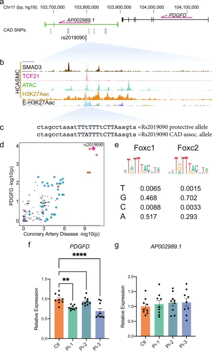
We map allele-specific transcription factor binding, chromatin accessibility and 3D chromatin looping in primary human coronary artery smooth muscle cells, and combine these molecular QTLs with CRISPR interference to connect risk variants to their target genes. This has revealed causal mechanisms at loci including TCF21, SMAD3, ZEB2, PDGFD, FN1 and 9p21.3.
Key papers
Smooth muscle cell fate in atherosclerosis
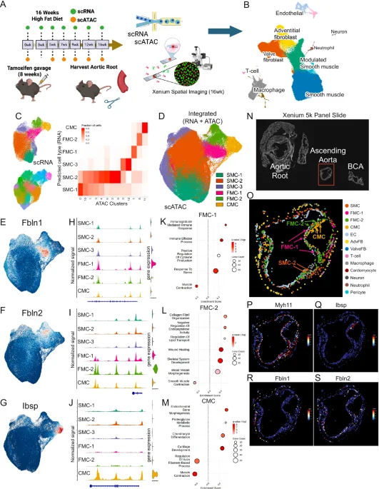
Smooth muscle cells (SMCs) are the cell type in which CAD genetic risk is most strongly enriched. During atherosclerosis they undergo "phenotypic modulation" into fibromyocytes that stabilize the fibrous cap, or chondromyocytes that drive calcification. Using lineage tracing with single-cell genomics, we discovered that the CAD gene TCF21 promotes the protective transition, and that SMAD3, ZEB2, AHR and PDGFD control the balance between fates.
Our dense single-cell RNA and chromatin timecourses now trace these trajectories step by step, identify the transcription factor networks that drive them, and show where along each trajectory inherited risk acts.
Key papers
Single-cell and spatial atlases of human arteries
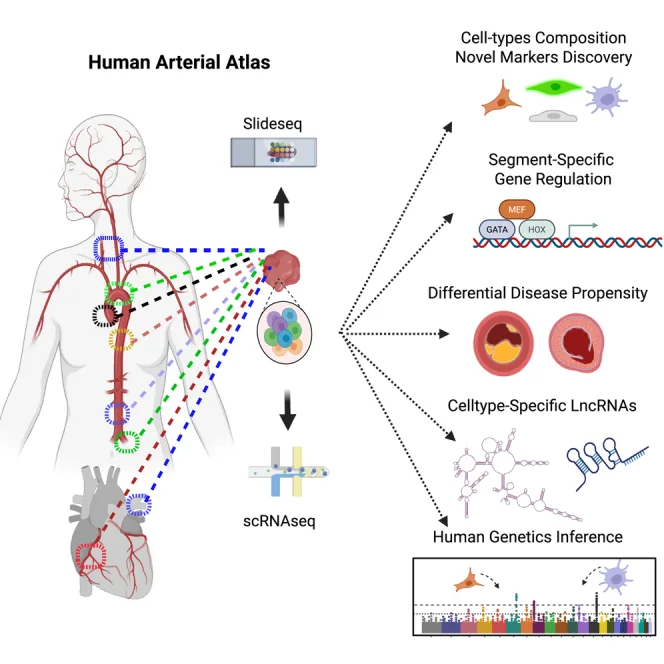
As part of the Chan Zuckerberg Initiative Human Cell Atlas, we built a single-cell and spatial atlas of healthy human arteries across multiple segments — from the aortic root and coronary arteries to the carotid, pulmonary and iliac arteries. Arterial smooth muscle cells, fibroblasts and endothelial cells carry segment-specific programs that reflect their embryonic origin and help explain why disease strikes some vessels and spares others.
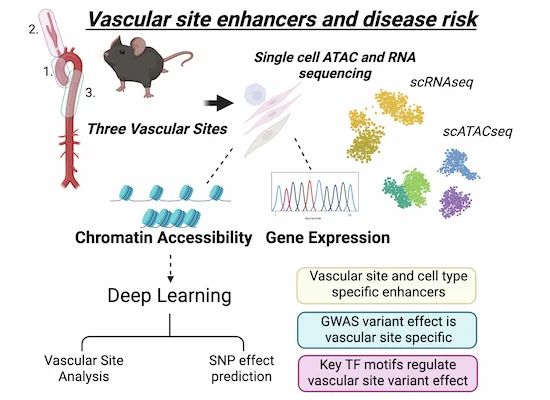
Paired single-cell chromatin maps reveal vascular site–specific enhancers, and deep-learning models of chromatin accessibility (ChromBPNet) predict how disease variants act in each cell type and vascular site.
Key papers
Functional genomics at scale
To move from single loci to the whole genetic architecture of CAD, we perturb hundreds of candidate genes and enhancers at once in human coronary artery smooth muscle cells and in mouse arteries, reading out each perturbation by single-cell RNA sequencing.
As part of the NHGRI Impact of Genomic Variation on Function (IGVF) consortium we have generated CRISPR interference screens with three complementary designs: Parse Perturb-seq targeting the promoters of genes at CAD loci, TAP-seq targeting candidate cis-regulatory elements across CAD loci with a targeted readout of nearby genes, and in vivo Perturb-seq in Myh11-Cre; Rosa26-LSL-dCas9-KRAB mice, in which an AAV guide library silences candidate genes specifically in aortic smooth muscle cells. Released datasets are available on the IGVF Data Portal, with more to follow.
37 released CRISPRi single-cell datasets
Additional datasets are in processing and will be released through the portal.
Browse on data.igvf.org →- 19Parse Perturb-seqCRISPRi of transcription start sites of genes at CAD loci in HCASMC-hTERT cells, scRNA-seq readout.
- 14TAP-seqCRISPRi of candidate cis-regulatory elements across CAD loci, targeted scRNA-seq readout.
- 4in vivo Perturb-seqAAV guide library against 19 genes in Myh11-Cre; Rosa26-LSL-dCas9-KRAB mouse aortic SMCs.
Key papers
Wet lab and dry lab, together
Research in the lab is carried out with both bioinformatic and experimental approaches. Some trainees focus on one discipline, but we encourage everyone to gain expertise in both generating and analyzing the high-dimensional datasets that reflect the transcriptional and epigenetic mechanisms of complex human traits.
- IGVF Consortium — With five other Stanford labs, decoding how cardiovascular GWAS variants affect genome function and disease.
- CZI Human Cell Atlas — Single-cell transcriptomic and epigenomic features of human arterial segments, released openly on CELLxGENE.
- ATHENA Network — A transatlantic collaborative laboratory (Stanford, Cambridge, Karolinska, Helsinki, WashU) to validate causal atherosclerosis genes.